Pory 78, Warsaw, Poland
mapDiagnostic Imaging Centre
Pory 78, Warsaw, Poland
mapWe cooperate with international insurers. Check if we accept your insurance.
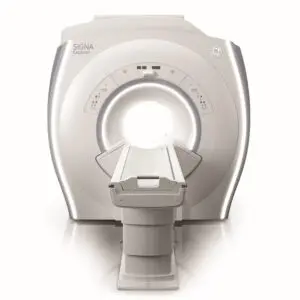
Magnetic resonance imaging (MRI) is a very accurate method of imaging mainly the soft tissues of the human body, including those located deep in the body that are therefore not visible in, for example, ultrasound examination (US). MRI is based on the phenomenon of nuclear magnetic resonance and does not use X-rays. It involves taking many scans of the examined organ, which allows the structure to be, as it were, “sliced” into thin sections. In each scan (sequence), many slices are taken in different planes, and, depending on the indications and on the area examined, the slices can be thicker or thinner.
Learn more about magnetic resonance imaging: https://carolina.pl/diagnostyka/rezonans-magentyczny/
We perform magnetic resonance examinations in both children and adults.
Magnetic resonance imaging at Carolina Medical Center is used for:
We also perform arthrography, an examination carried out after intra-articular administration of 10–20 ml of contrast agent or physiological saline. The joint puncture is performed immediately before the MRI examination in our laboratory under ultrasound guidance. The joint becomes distended, which improves imaging of the examined area, because the contrast flows into injuries/fissures in the cartilage, tendons or labrum. After the examination the joint cavity returns to its original size.
Arthrography is used to assess damage to the cartilaginous and ligamentous elements of joints with more complex structures, such as the shoulder, hip, wrist and, less frequently, the knee.
We also offer examinations with intravenous contrast.
Magnetic resonance imaging is used as a complement to other imaging examinations, which in some situations may be insufficiently precise. MRI is the only imaging examination that enables a reliable assessment of the healing of the cruciate ligament and the meniscus after surgery.
The MRI examination is painless but requires the patient to remain motionless for more than an hour. The examination does not require any special preparation.
Before the examination, please inform us about:
Required medical documentation:
Clothing and make-up:
For the examination, you will be asked to leave your clothing in the changing room. Only underwear may remain, provided it contains no metal elements (e.g. underwires in bras). You will receive a disposable non-woven gown and foot covers from us.
All electronic and magnetic items (e.g. payment cards), items containing metal and jewellery must be left in the changing room for the duration of the examination – please keep this in mind when planning your visit.
When coming for the examination (especially a head scan), you should avoid wearing make-up and hair spray – cosmetics contain trace metals which may affect the image.
Eating and drinking before the examination:
It is recommended that you do not drink large amounts of fluids. You do not need to fast before the examination.
EXCEPTION: In the case of an examination with intravenous contrast, please fast before the examination.
The examination is non-invasive and completely painless; however, for many patients the prospect of lying motionless in a narrow and noisy MRI tube is very stressful. Find out how the examination proceeds and what to expect, so nothing takes you by surprise.
Before the examination you will be given a questionnaire to complete, in which you will be asked not only for your personal details but also for information about your state of health. You will also need to give written consent to the examination. Filling in the documentation takes about 15 minutes, so please arrive at the clinic in advance.
In some cases a special contrast agent is administered intravenously to the patient during the examination. In examinations with intravenous contrast, a cannula will be inserted. You will then be asked to lie in the appropriate position, depending on the examination, on the sliding table in the centre of the MRI scanner. To increase your comfort, the staff may provide you with a blanket and a pillow.
Throughout the examination you must not move, so that the image is not distorted. The tunnel is fitted with air conditioning, lighting and monitoring, thanks to which the staff will be in constant contact with you and can respond to any signals from you. In case of any sudden deterioration of your wellbeing during the examination, report it to the staff by squeezing the alarm bulb that you will hold in your hand throughout the examination.
Each sequence of the magnetic resonance examination lasts from 1 to 8 minutes. During this time you will hear a loud knocking sound of intensity similar to a running washing machine. Before the examination begins, the staff will provide you with protective headphones or earplugs. During the examination you can also listen to music or a radio programme of your choice – our laboratory is equipped with a modern, special sound system connected to an iPad.