Stress X-ray

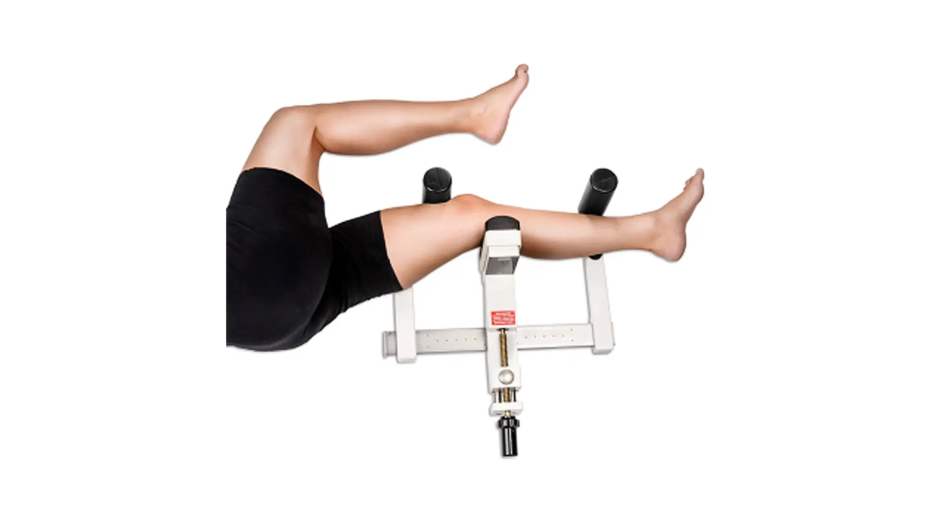
Stress X-ray with the use of the TELOS device is a test performed during the diagnosis of joint instability. It allows for precise and objective determination of whether the tested ligament is healthy (whether it fulfills its function after an injury or surgery). The test is performed in a forced (stress) position, and the assessment is usually comparative – we examine the healthy side and the sick side.

Stress images are taken:

  • in the case of suspected ligament insufficiency and ambiguous results of other tests (imaging and orthopedic),
  • to evaluate the results of surgical treatment.

In the Carolina Medical Center, stress X-rays are used to examine the ankle joint, knee joint and elbow joint.

The examination is performed on the basis of a referral.

Uwaga! Wymagane skierowanie
Umów się na wizytę

Knee-joint:

  • anterior cruciate ligament (the so-called anterior drawer)
  • posterior cruciate ligament (the so-called posterior drawer)
  • tibial collateral ligament,
  • fibular collateral ligament.

Elbow joint:

  • collateral ligaments.

Ankle:

  • medial ligament,
  • anterior talofibular ligament,
  • anterior drawer of the ankle.

An example of an image made in a stress X-ray: Knee joint in lateral view.