Magnetic resonance imaging

Magnetic resonance imaging (abbreviated as MRI) is a very accurate method of imaging mainly soft tissues of the human body, including those deeply located, and therefore not visible, for example, in an ultrasound examination. Magnetic resonance imaging is based on the phenomenon of a magnetic field and does not use x-rays. It consists in performing many sequences of the examined organ, and similarly to computed tomography examination, it allows to “cut” the examined structure into slices. There are many slices in each sequence, and depending on the indications and the area to be examined, the slices may have a greater or lesser thickness.

High quality examinations

At the Carolina Medical Center, the MRI examination takes longer than in other facilities because we perform more sequences of the examined organ, and our images have a very high resolution. In other places, a standard examination of, e.g., a knee joint takes about 30 minutes, while in our facility it takes up to over an hour. This guarantees us and our patients a more accurate diagnosis and gives a better chance of recovery.

An example of an MRI examination performed at the Carolina Medical Center in Warsaw and another medical facility:

Images of the knee joint – damage to the meniscus and cartilage (MRI)

Uwaga! Wymagane skierowanie.
Umów się na wizytę

Fig. 1 – examination performed at the Carolina Medical Center

Fig. 2 – examination performed at another medical facility

The tests are performed under the supervision of doctors – radiologists who specialize in orthopedic diagnostics.

The Imaging Diagnostics Center uses the General Electric Signa 1.5T HDxt Adv, equipped with a set of coils dedicated to orthopedic examinations, adapted to joint examinations, which further increases the precision of the diagnosis. The device is also equipped with a whole-body diffusion option designed to detect inflammatory lesions or diseased areas.

The scope of MRI examinations performed at the Carolina Medical Center

We perform magnetic resonance imaging in both children and adults.

Magnetic resonance imaging at the Carolina Medical Center is used for:

  • studies of the musculoskeletal system: joints, tendons and muscles.
  • detecting inflammatory lesions, overload changes or muscle swelling at an early stage (before the onset of ailments), which is extremely important, especially for people who play sports.

We also carry out arthrography, which is an examination performed after the intra-articular administration of 10-20 ml of a contrast agent or saline. Puncturing of the joint is performed immediately before the MRI examination in our facility under the control of the ultrasound image. The joint becomes distended, which promotes better imaging of the examined area, because the contrast affects the damage/fissures of cartilage, tendons, and the labrum. After examination, the joint cavity returns to its original size.

Arthrography is used to assess damage to the cartilage and ligamentous elements of more complex joints, such as the shoulder, hip joint, wrist and, more rarely, the knee joint.

Magnetic resonance imaging is used as a supplement to other imaging tests, which in some situations may be too inaccurate. MRI, as the only one of all imaging tests, allows for a reliable assessment of the healing of the cruciate ligament and meniscus after surgery.

Preparation for magnetic resonance imaging

MRI examination is painless, but requires staying still for up to over an hour. The examination does not require any special preparation.

Before starting the test, inform about:

  • medications you are taking – the doctor who will refer you to an MRI scan will decide whether you can take them before and after the examination,
  • presence in the body of metal implants (vascular clips, prostheses, screws, wires, plates). In this case, it is necessary to present a document / certificate containing information / characteristics of the alloy from which the implant is made or a certificate of admission to testing in a magnetic field – issued by the manufacturer. Without these documents, the test cannot be performed for the sake of your safety.
  • presence of other metallic bodies in the body (shards, bullets, etc.)
  • possible pregnancy – MRI is not recommended in the first trimester of pregnancy.

Required medical documentation:

  • A medical referral is NOT required to undergo an MRI scan.
  • Be sure to bring previous results of the examined body part with you, if you have such results.
  • If, during the examination, a contrast agent is administered intravenously or intraarticularly, the current result of the creatinine level in the blood should be presented – the validity period of the test is 1 month.
  • Remember to also bring your ID card!

Clothes and makeup:

You will be asked to leave your clothes in the changing room during the examination. Only underwear is not removed, as long as it does not have metal elements (e.g., underwire in a bra). You will receive a disposable t-shirt and foot pads. All electronic and magnetic items (e.g., payment cards) containing metal and jewelry must be left in the changing room during the examination – remember about this when planning your visit.

When going for an examination (especially in the case of a head examination), you should not apply makeup and hairspray – cosmetics contain particles of non-ferrous metals, which may affect the image.

Consuming drinks/food before the test:

It is recommended not to drink large amounts of fluids. You do not need to be on an empty stomach when you start the test..

EXCEPTION: In the case of a planned examination with intravenous contrast, we ask the patient to be on an empty stomach.

What happens during an MRI scan?

The examination is non-invasive and completely painless, however, for many patients the prospect of being motionless in a narrow MRI tube is associated with a lot of stress. Find out how what happens during the examination and what to expect so that nothing surprises you.

Before the examination, you will receive a questionnaire to fill in, in which you will be asked not only for personal data, but also information about your health condition. You will also need to give your consent in writing so that the examination can be performed. Filling out this documentation will take you about 15 minutes, so please arrive at the clinic in advance.

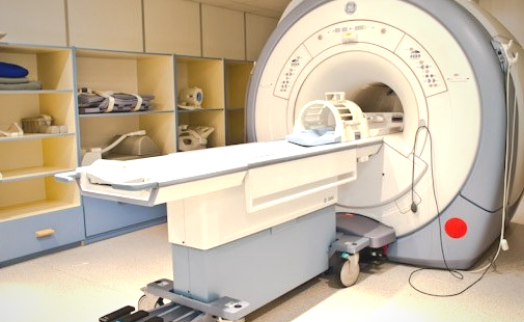
In some cases, during the examination, the patient is administered a special contrast agent intravenously. For tests with the intravenous administration of a contrast agent, a cannula will be inserted. Then you will be asked to lie down in the appropriate position, depending on the examination you are doing, on the extendable table in the center of the MRI scanner. In order to increase your comfort, the staff can provide you with a blanket and a pillow.

Do not move during the entire duration of the examination so as not to distort the image. The tunnel is equipped with air conditioning, lighting, and monitoring thanks to which the staff will be in constant contact with you and will be able to react to any signals from your side. If you suddenly feel unwell during the examination, report it to the staff by pressing the button on the signaling device, which you will hold in your hand throughout the examination.

Each MRI scan sequence takes 3 to 10 minutes. During this time, you will hear a loud knocking sound similar in intensity to a spinning washing machine. Before starting the examination, the staff will provide you with noise-reducing headphones (or earplugs for head and spine examinations). In a situation where several images need to be taken, the table automatically moves to the appropriate position, but the patient remains motionless the whole time.

Contraindications for MRI examination:

  • pacemaker,
  • cochlear implants,
  • ferromagnetic vessel clips,
  • infusion pump,
  • the presence of a metallic foreign body in the eye,
  • pregnancy (1st trimester).

MRI results

You can receive the MRI images immediately after its completion on a CD/DVD – report to the ER reception desk on the ground floor to collect the CD. The test description can be collected after 6 working days after receiving an SMS notification.

  • You can personally collect the description on the 2nd floor in the Medical Documentation Department, from Monday to Thursday, from 08:00 am to 7:00 pm, and on Fridays from 08:00 am to 6:00 pm.
  • If you would like to receive the description (or images) by e-mail or post, report to the employee of any reception desk during the registration of the examination or after its completion. You can also request this by phone after completing the survey.