Pory 78, Warsaw, Poland
mapDiagnostic Imaging Centre
Pory 78, Warsaw, Poland
mapWilenska 44, Gdansk, Poland
mapWe cooperate with international insurers. Check if we accept your insurance.
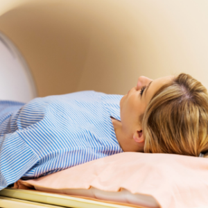
Computed tomography (CT) is a diagnostic method that uses X-rays to obtain cross-sectional images of the inside of the body.
The main components of a CT scanner are an X-ray tube and detectors mounted on opposite sides of a circular gantry. The X-ray tube generates radiation that passes through the body of the patient lying inside the scanner and is received by the detectors. The X-ray beam passing through the body is absorbed to different degrees by different tissues. The detectors measure the level of X-ray absorption, and on this basis a cross-sectional image of the body is created. Bones, which absorb radiation most strongly, appear white, while other tissues appear in various shades of grey.
Computed tomography is characterised by high spatial resolution. Two- and three-dimensional images of the examined structures can be obtained.
Computed tomography is used to diagnose complex bone injuries that are difficult to detect on X-ray, such as:
CT examinations are also performed on patients with contraindications to MRI (e.g. patients with metal implants, cardiac pacemakers, etc.).
We also perform CT arthrography examinations.
Learn more: https://carolina.pl/centrum-diagnostyczne/badanie-rentgenowskie/