Pory 78, Warsaw, Poland
mapDiagnostic Imaging Centre
Pory 78, Warsaw, Poland
mapWe cooperate with international insurers. Check if we accept your insurance.
An X-ray examination, also colloquially known as a radiograph, is an imaging technique that uses X-rays. An X-ray examination is the basic method of imaging the skeletal system thanks to its wide availability, relatively low cost and short examination time. X-rays have been used in medicine for over 100 years, making them a very well-known and safe technique.
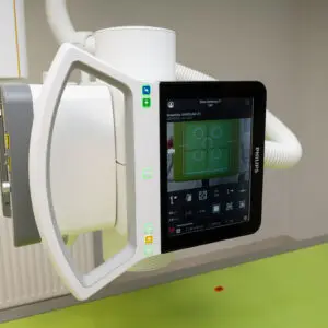
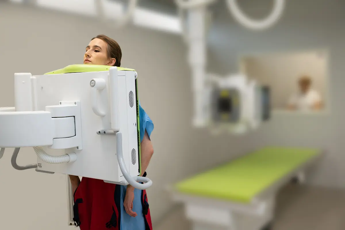
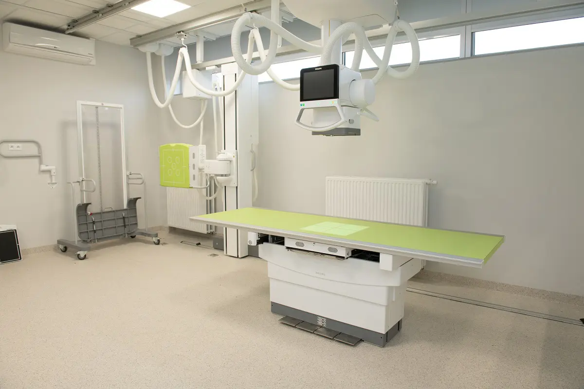
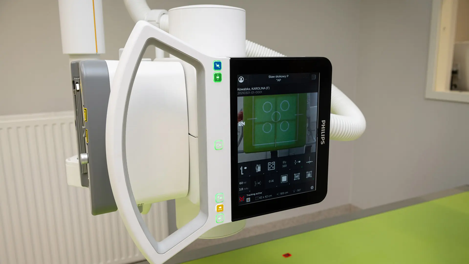
Carolina Medical Center uses two modern X-ray units for X-ray examinations. Both units are configured to make the examination as easy as possible for patients, and through the use of modern digital techniques they also reduce the radiation dose received.
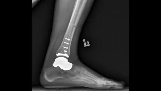
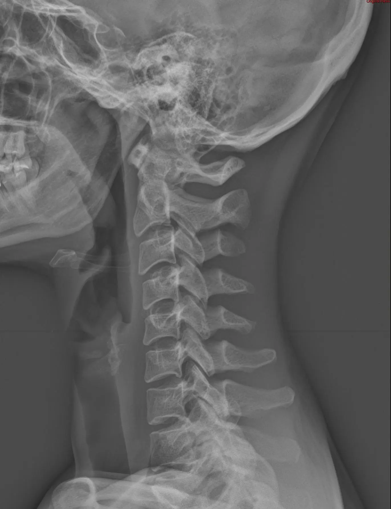
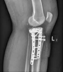
The X-ray image is used to assess the structure of bone tissue – for example, possible cracks and fractures. In most patients with bone injuries, X-ray imaging makes it possible to plan treatment and monitor healing progress. X-rays are not suitable for assessing other tissues, such as injuries to ligaments or muscles. An X-ray examination shows only the outline of soft tissues, not their structure.
The X-ray unit at Carolina Medical Center is used for:
At our clinic we perform the full range of X-ray examinations used in the diagnosis of musculoskeletal conditions. We also perform full-length (postural) X-rays of the lower limbs, covering the entire lower limbs, including the hip, knee and ankle joints. This examination allows the assessment of axial disorders or leg length asymmetry.
We very often perform comparative X-rays of the limbs, which significantly increases examination effectiveness and helps detect pathologies, especially in our youngest patients.
Learn more: https://carolina.pl/zespol/rentgen/