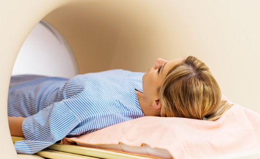
Computed tomography

Computed tomography (CT) is a diagnostic method that uses X-rays to obtain cross-sectional images of the inside of the body.

The main elements of the CT scan are the X-ray tube and detectors which are suspended on opposite sides of a circular rim. The X-ray tube generates radiation that passes through the body of the patient lying inside the tomograph and is picked up by detectors. The beam of X-rays penetrating the body is absorbed to varying degrees by the tissues in the human body. The detectors are able to measure the level of absorption of X-rays and on this basis a section of the body is created. Bones, which absorb radiation most strongly, will be visible in white, and other tissues in various shades of gray.

Computed tomography is characterized by high spatial resolution. It is possible to obtain two- and three-dimensional images of the examined structures.

At the Carolina Medical Center, we use the Philips Brilliance CT 40.

Indications for computed tomography in the Department of Diagnostic Imaging of the Carolina Medical Center

Computed tomography is used to diagnose complex bone lesions that are not easy to detect in X-ray examination:

  • to visualize and accurately assess complex fractures before the planned surgery,
  • assess postoperative complications (e.g, assess bone union disorders, inflammatory changes, destabilization of the anastomosis),
  • assess bone fixation,
  • assess the correct alignment of bone structures,
  • assess acquired or congenital bone deformities,
  • assess extra-articular calcifications,
  • assess limb/bone rotation, assess bone structure changes – inflammations, tumors,
  • assess intra-articular structures after intra-articular administration of a contrast agent – CT arthrography.

Patients with contraindications to MRI examination can undergo CT examination (patients with metal implants, pacemakers, etc.).

We also carry out computed tomography arthrography.

Arthrograph is an examination performed in radiology of the musculoskeletal system, consisting in the administration of 10-20 ml of a contrast agent by intra-articular injection. The joint puncture is performed immediately before the tomography, in our laboratory using ultrasound imaging. The joint becomes distended, which promotes better imaging of the examined area, because the contrast affects the damage/fissures of cartilage, tendons, and the labrum. After examination, the joint cavity returns to its original size.

Arthrography is used in the assessment of the labrum of the shoulder joint, the labrum of the hip joint, partial injuries of tendons and ligaments, injuries of the hand ligaments, postoperative injuries of tendons and menisci, and surgical planning.

This examination is only performed in patients over 18 years of age.

Preparation for computed tomography

Computed tomography examination is painless, but requires staying still for a relatively short time (up to 10-15 minutes). The examination does not require any special preparation.

Before starting the test, inform about:

  • allergies and allergic reactions, including contrast agent allergy,
  • bleeding tendency, pregnancy or suspected pregnancy,
  • claustrophobia,
  • medications you are taking – the doctor who will refer you to a CT scan will decide whether you can take them before and after the examination,
  • the presence of foreign elements in the body.

Required medical documentation:

A medical referral is required to undergo a CT scan. If, during the examination, a contrast agent is administered intravenously or intraarticularly, the current result of the creatinine level (eGFR) in the blood should be presented – the validity period of the test is 1 month. If you have already had a computed tomography or other diagnostic tests (X-ray, CT, MRI) of the examined anatomical area, please bring the previous results with you (description + plates). Remember to also bring your ID card!

Clothes

You will be asked to leave your clothes in the changing room during the test. Only underwear is not removed, as long as it does not have metal elements (e.g., underwire in a bra). You will receive a disposable t-shirt and foot pads.

Remember to remove all metal objects: jewelry, glasses, external hearing aid – if they are within the examined area.

Consuming drinks/food

In the case of tests with a contrast agent, you should come on an empty stomach – you should not eat food at least 6 hours before the examination. After the examination is completed, you can eat. After the test, in order to facilitate the removal of the contrast agent from the body, it is recommended to drink plenty of fluids (about 2.5 liters of still water or unsweetened drinks in the next 24 hours).

Contraindications for CT examination:

Computed tomography uses X-rays to obtain images of a cross section of the body. Radiation doses during a CT examination can be up to 500 times higher than during a regular X-ray image. Therefore, it cannot be carried out too frequently and at too short intervals. In children, tomography is limited to the necessary minimum.

Pregnancy is an absolute contraindication – a large dose of X-rays could be harmful to the fetus. The test can be performed at any time during the menstrual cycle, but it is recommended to perform the test up to the 10th day of the cycle to avoid exposure to radiation in the case of an early, undiagnosed pregnancy.

Attention! Referral required.
Book an appointment over the phone

Warszawa (Mokotów)

+48 22 35 58 200

Warszawa (Żoliborz)

+48 22 25 00 500

Contraindications to the administration of iodine contrast agent:

Iodine contrast agents are most often administered intravenously in computed tomography, but they can also be administered by other routes, e.g., intra-articular. These agents enhance the contrast of the image due to their different absorption by different tissues.

Absolute contraindications for contrast medium administration include documented, severe hypersensitivity to iodine contrast agent, renal failure, and overt hyperthyroidism.

How is a computed tomography examination performed?

Before the examination, you will receive a questionnaire to fill in, in which you will be asked not only for personal data, but also information about your health condition. Filling out this documentation will take you about 10 minutes, so please arrive at the clinic in advance.

Computed tomography is a painless and short examination. Depending on the examined organ, it takes from a few seconds to a few minutes.

If a contrast agent is administered intravenously for examination, it is necessary to penetrate the vein and insert a cannula. Occasionally, after administration of a contrast agent, the patient may experience a temporary feeling of warmth or pressure on the bladder, possibly accompanied by slight nausea. Side effects are rare and usually short.

The medical staff will ask you to lie down on a special movable table, which will be inserted into the device during the examination. Depending on the type of examination, you will be asked to lie down on your stomach, back or side. The X-ray tube will rotate around you and capture images.

You should stay still during the examination, as movements may blur the image. You may be asked to hold your breath for a few moments.

After scanning the body in the selected range, the platform will leave the tomograph and the examination will be completed.

The next stage is the analysis of the examination performed by the doctor, digital processing of the obtained images, and then preparation of the description and documentation of the examination.

CT scan results

You can receive the CT scan pictures 30 minutes after the examination is completed on a CD/DVD – report to the ER reception desk on the ground floor to collect the CD. The test description can be collected after 6 working days after receiving an SMS notification.

  • You can personally collect the description on the 2nd floor in the Medical Documentation Department, from Monday to Thursday, from 08:00 am to 7:00 pm, and on Fridays from 08:00 am to 6:00 pm.
  • If you would like to receive the description (or images) by e-mail or post, report to the employee of any reception desk during the registration of the examination or after its completion. You can also request this by phone after completing the survey.