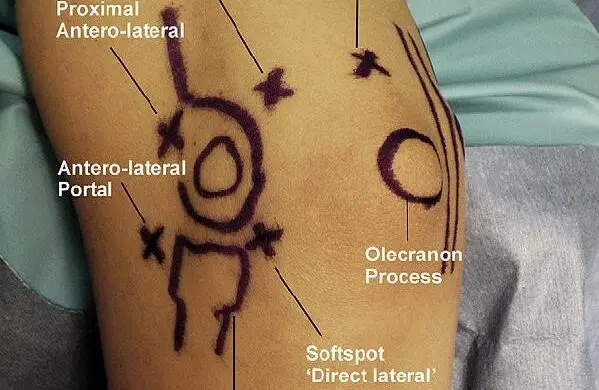
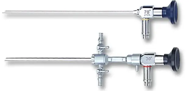
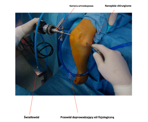
An arthroscopy is a minimally invasive surgical procedure involving implementation of a camera as well as surgical instruments to the joint. Surgical instruments are implanted via small skin cutting that measures approximately 0.5 cm (portals). The number of portals and their placement depend on the procedure we want to perform. It can be the arthroscopy of a front or rear portion. The joint is filled with physiological salt and camera generated image is transferred to the high-resolution monitor. An operator is conducting this procedure, looking at the onscreen image.
It is a highly modern surgical method, although the first arthroscopy of the elbow-joint was conducted as early as 1931, by Burman. During last two decades the surgical instruments have been strongly developed, as well as a technique. Improving the scope of the procedure and number of indications for the arthroscopy.
The possibilities that this method creates are unimaginable. Many stressful open surgeries were ousted by this perfect technique. Huge amounts of publications in Polish and international literature, as well as our own experience show the advantage of this technique. The number of complications from an open surgery is drastically decreasing, and the treatment result is usually better then with traditional methods.
Furthermore, it is a surgical procedure and as any surgical procedure it can cause complications, like nerve damage, inflammation etc. Conducting this procedure by an experienced surgeon utilizing the most up to date equipment and within the operating block significantly decrease number of this occurrences.
Elbow-joint arthroscopy – indications
- Emoval/replantation of loose bodies from an elbow-joint
Loose bodies – bone or chondral elements – are usually the result of injury/fracture within the joint. Those bodies can limit movement range and damage the joint. They can also be a very important element of intra-articular structures, e.g. cartilage fragment within loaded area that must be replanted. - Femoval of scars and adhesions from a joint. They usually form as a result of an injury and internal joint bleeding, like posttraumatic arthrofibrosis.
- Removal of bone adenoids/osteophytes which form as a result of chronic overloads, like thrower’s elbow, injuries or osteoarthritic changes.
- Mobilization of a movement range of a joint related to the joint capsule contracture.
- Treatment of diseases and damages of joint cartilage, e.g. osteochondritis dissecans.
- Treatment of elbow-joint instability.
- Treatment of tennis elbow– working out a damage spot of the insertions of the wrist extensors muscles.
- Removal of the inflammatory changed synovium formed as a result of chronic rheumatoid-based disease.
- Treatment of an elbow-joint fractures, , e.g. the coronoid process fractures.
- Release of nerves incarcerated in scars.
- Treatment of intra-articular inflammations.
Elbow-joint arthroscopy – przebieg zabiegu
Anaesthesia: Usually it is the anaesthesia of a brachial plexus, meaning that only operated limb is anaesthetized. Patient can consciously participate in the procedure.
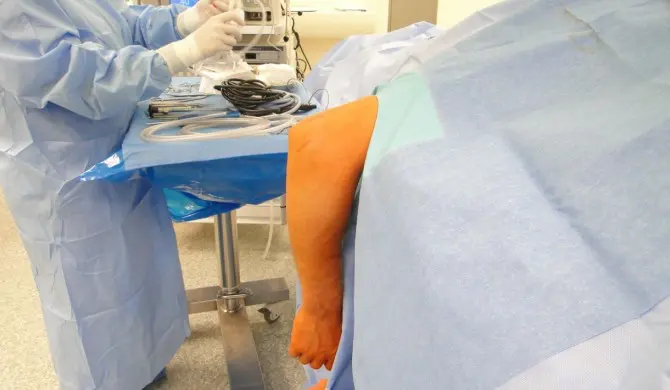
Position: Patient lays on his stomach. Operated limb is positioned on a special underlay that allows free bending and straightening of an elbow-joint.