Ankle arthroscopy
A minimally invasive method of joint operation, used also in case of ankle injuries.
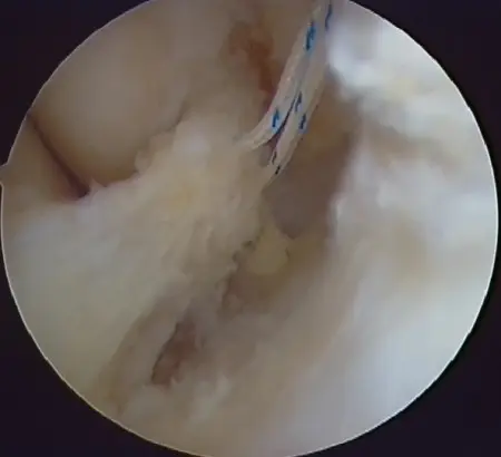
Arthroscopy is not such a novelty – first arthroscopy was performed nearly 100 years ago in 1918! In those days it was treated mainly as a diagnostic method that allowed looking into a joint as if through a keyhole. Currently, it is performed mainly as a treatment method, that allows performance of many procedures within a joint, including ligaments reconstruction and cartilage correction.
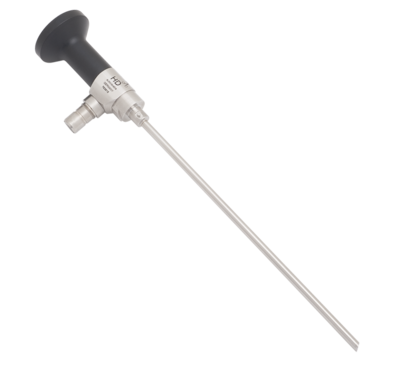
Arthroscopy advantage over traditional operating methods is that there is no need for full joint opening. Usually few (2-3) small (0.5 cm) cuts is enough. Through those cuts a video camera and tools are introduced. Thanks to these actions, coming back to health can be much faster, scars fully invisible and joint surrounding tissue damage – minimal.